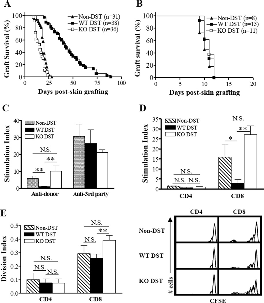
Figure 1.
CD47 expression on donor cells is required for prolongation of donor skin grafts by DST. (A,B) Donor (B6; A) and third-party (B10.A; B) skin allograft survival in naive bm1 mice (Non-DST) and bm1 mice that received DST (i.e., 1×10⁷ splenocytes) from WT (WT-DST) or CD47 KO (KO-DST) B6 donors 7 days prior to skin grafting. Results in (A) and (B) are the combined data of 7 and 2 independent experiments, respectively. (C, D) In vitro MLR. Total and purified CD4+ or CD8+ T cells were prepared from recipient splenocytes at day 7 post-DST, and cocultured with equal number of irradiated (30 Gy) splenocytes (stimulators) from B6 (donor) or B10A (3rd-party) mice. Results (mean±SDs) are presented as stimulation index and the data of 2 independent experiments are combined (n=5 per group). Shown are anti-donor and anti-3rd-party MLR of total T cells (C) and anti-donor MLR of purified CD4+ and CD8+ T cells (D). (E) In vivo MLR. Splenocytes were harvested from non-DST controls (n=5) and the recipients of WT (n=9) or CD47 KO (n=5) DST 7 days after DST, labeled with CFSE, and adoptively transferred into lethally irradiated (9.5 Gy) B6/Ly5.2 (CD45.1) congeneic mice. Proliferation of injected bm1 CD4+ and CD8+ T cells was measured by flow cytometry 3 days after adoptive transfer. Results from 2 independent experiments are combined. Shown are division index (left; mean±SDs) and flow cytometry profiles (right) of bm1 CD4+ (i.e., CD45.1−CD4+) and CD8+ (i.e., CD45.1−CD8+) T cells. *P<0.05; **P<0.01; N.S., not significant.

