
Fig. 2.
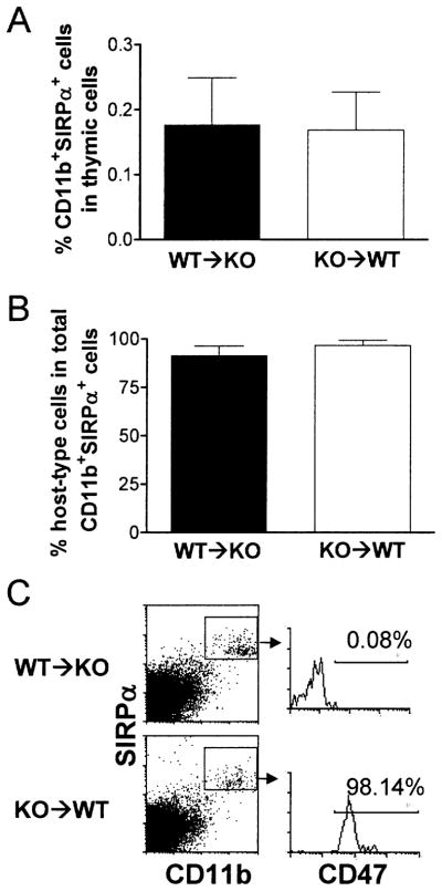
Repopulation of thymic grafts by recipient macrophages. Thymic grafts were harvested from WT thymus-grafted CD47 KO mice (WT → KO; n = 6) and CD47 KO thymus-grafted WT mice (KO → WT; n = 5) 7 weeks after thymic transplantation, and single cell suspensions were prepared and analyzed for SIRPα+CD11b+ macrophage repopulation. Donor- and recipient-derived cells were distinguished by staining with anti-CD47 mAb, in which recipient cells from WT → KO and KO → WT mice were CD47− and CD47+, respectively. Shown are percentages (mean ± SEM) of SIRPα+CD11b+ macrophages in total thymic cells (A), percentages (mean ± SEM) of host-type SIRPα+CD11b+ macrophages in total macrophages (B), and representative staining profiles (C).