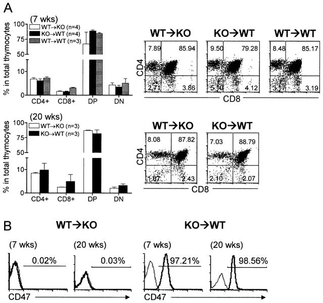
Fig. 3.
Phenotypic analysis of thymocytes in thymic grafts. Thymic grafts were removed from WT thymus-grafted CD47 KO mice (WT → KO) and CD47 KO thymus-grafted WT mice (KO → WT) at weeks 7 and 20 and from WT thymus-grafted WT mice at week 7 after thymic transplantation, and analyzed by flow cytometry. (A) Percentages (mean ± SEM) of CD4+, CD8+, CD4+CD8+ (DP), and CD4−CD8− (DN) cells (left panel) and representative flow cytometric profiles (right panel). (B) Representative profiles of anti-CD47 (thick line) and isotype (thin line) antibody staining. Recipient-derived cells in the grafts from WT → KO and KO → WT mice are CD47− and CD47+, respectively.

