Figure 5.
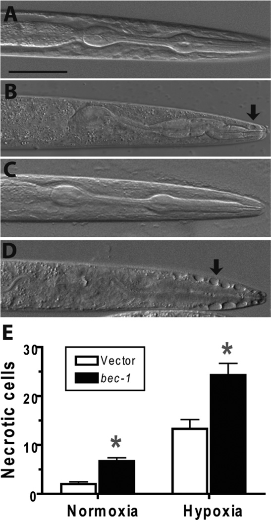
Effect of bec-1 knockdown on necrotic pathology. Wild type larval animals with vector or bec-1 RNAi treatment underwent a twelve hour hypoxic or normoxic incubation then scored after recovery for myocyte number. Necrotic cells were visualized and counted using Nomarski optics. Cells with markedly swollen morphology and lack of nuclei were scored as necrotic. (A) Normoxic vector-treated animal showing normal cellular morphology in the pharyngeal region. (B) Hypoxic vector-treated animal showing a few necrotic-appearing cells (arrow). (C) Normoxic bec-1(RNAi) animal showing normal cellular morphology. (D) Hypoxic bec-1(RNAi)-treated animal showing multiple necrotic cells adjacent to the anterior pharynx (arrow). (E) Comparison of necrotic cell number in bec-1(RNAi) vs vector-treated animals after normoxic or hypoxic incubation. Data are mean ± sem of 35 animals/condition. * - p < 0.01 vs vector control. Scale bar = 20 µm.

