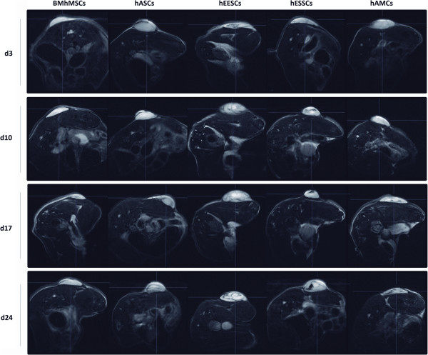
Figure 2.
In vivo xenograft imaging of mice using a magnetic resonance labeling (MRI) scanner. Scans were performed at 3, 10, 17, and 24 days after intravenous injection of super-paramagnetic iron oxide (SPIO)-labeled mesenchymal stem cells (MSCs). Tumors were visible as lighter areas in transverse sections. The recruitment of SPIO-labeled MSCs to tumors resulted in decrease of signal intensity (SI) and the visualization of darker areas in tumor sites. The same animals are represented over the entire period.

