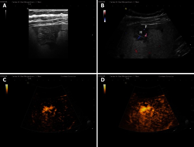
Figure 2.

Shunt hemangioma. Shunt hemangiomas are typically small (often < 20 mm) with abundant arterio(porto-)venous shunts (functionally described as high flow hemangiomas). They are often surrounded by less fat-containing hypoechoic liver parenchyma (A, B) due to the dominant arterial blood flow in comparison to the reduced portal venous perfusion. Arterial contrast enhancement of the shunt-hemangioma is also shown (C, D). H: Hemangioma; F: Less fat-containing hypoechoic area. Reproduced with permission from Dietrich et al[65].
