


Figure 3.
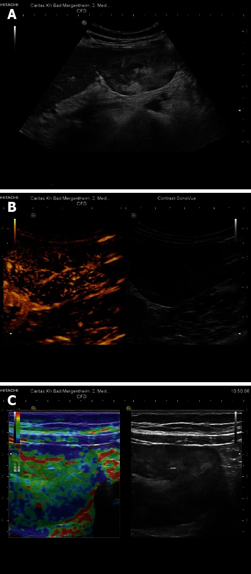
Teleangiectatic focal nodular hyperplasia. Pedunculated liver tumor, histologically teleangiectatic focal nodular hyperplasia with signs of peliosis. A: B-mode imaging shows heterogenous echogenicity; B: Contrast-enhanced ultrasound reveals central arterial blood supply; C: Real-time elastography shows harder periphery and softer central portions of the lesion.