Figure 3.

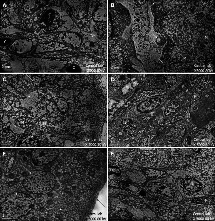
Transmission electron microscopy results showed. A: Transmission electron-micrograph of duodenum of rat of control group showing normal appearance of the core of the villi. Note eosinophil leucocytes (S) and close proximity of blood capillary (C) to the enterocyte basement membrane (BM); B: Transmission electron-micrograph of duodenum of breastfeeding rat group 1 showing local loss of microvillus border (long arrows) and swollen mitochondria with remnants cristae (M); C: Transmission electron-micrograph of duodenum of breastfeeding rat group 1 showing irregular nuclei (long arrow) with intercellular oedema (short arrow) and swollen mitochondria (M); D: Transmission electron-micrograph of duodenum of breastfeeding rat group 1 showing increased eosinophil leucocytes (S). Notice the nuclei of fibroblasts (F) and vacuolated enterocytes (V); E: Transmission electron-micrograph of duodenum of rat treated with human colostrum group 2 showing the enterocytes with vesicular nuclei (arrow) and regular microvillus border (black arrow). Notice the cell junction (head arrow)and abundant mitochondria(M); F: Transmission electron-micrograph of duodenum of rat treated with human colostrum showing normal appearance of the core of the villi with nuclei of lymphocyte (LY), nuclei of fibroblast (F) and eosinophil leucocytes (S).
