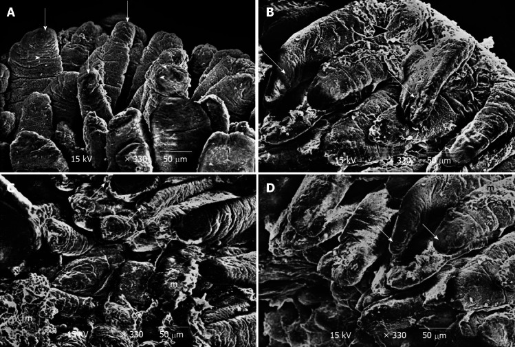
Figure 4.

Scanning electron microscopy results showed. A: Scanning electron-micrograph of duodenal villi of control group showing the villi with intact tips and border (short arrow). Notice the opening of goblet cells on the surface of the villi (arrow head); B: Scanning electron micrograph of duodenum of breastfeeding rat group 1 showing some of the villi are seen to be deformed (long arrow). Notice short villus (arrow head) is clearly seen; C: Scanning electron micrograph of duodenum of breastfeeding rat group 1 showing some villi with necrotic tips (short arrow). Notice distorted appearance of the other villi with accumulation of mucous (m); D: Scanning electron-micrograph of duodenum of rat treated with human colostrum group 2 showing duodenal villi with intact tips and borders (short arrow). Notice the presence of mucous on some villi (m).
