
Figure 1.
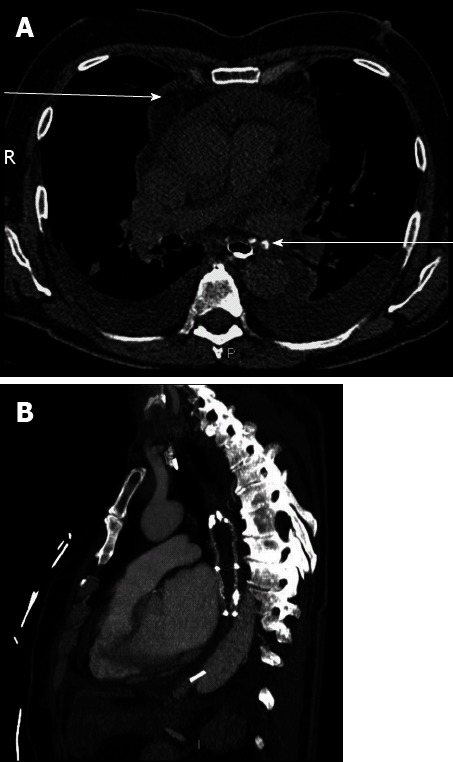
Computed tomography-scan. A: Computed tomography (CT)-scan with esophageal opacification. Pericardial effusion (upper arrow) and esophagopericardial fistula (lower arrow) were both present; B: CT-scan performed after esophageal stent placement and surgical drainage. Pericardial effusion was no longer present following esophageal stent insertion.