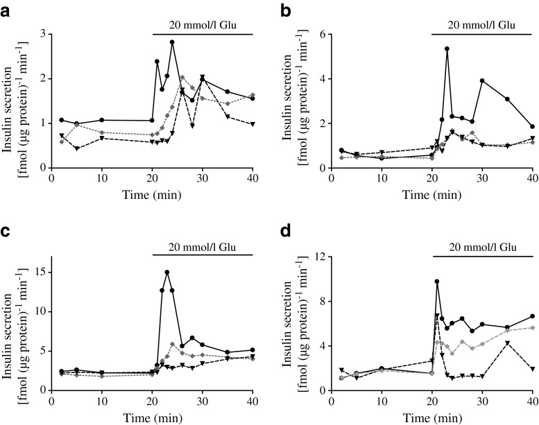
Fig. 5.
Glucose (Glu)-stimulated insulin secretion from MIN6 monolayers (a), pseudoislets (b), human islets (c) and mouse islets (d) in the absence (solid line) or presence of the PI3K inhibitors wortmannin (grey dotted line) or LY294002 (black dashed line). Insulin released to the medium was measured and normalised to protein. Results are representative of four or five separate experiments

