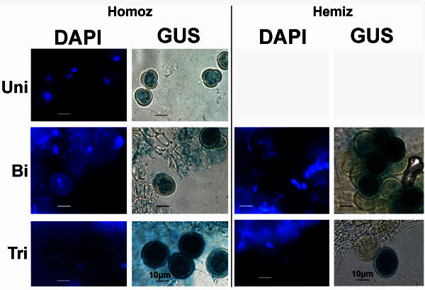
Figure 1.

Strong GUS staining in uninucleate microspores and pollen grains of thepAt5g17340:UidA:GFPmarker line. Strong GUS staining is detected in late uninucleate microspores (Uni), as well as in bicellular (Bi) and tricellular pollen (Tri) of plants homozygous for the pAt5g17340:UidA:GFP construct. Segregation of the marker gene (GUS stained and non-GUS stained pollen in a 1:1 ratio), as illustrated in the bicellular (Figure 1-Bi-Hemiz) and tricellular (Figure 1-Tri-Hemiz) pollen grains, indicates that the hemizygous plant has a single UidA marker gene insertion in the genome. Plates show DAPI stained nuclei (left column) of the GUS stained (right column) microspores and pollen grains in homozygous (Homoz) and hemizygous (Hemiz) plants. Uni- uninucleate microspore; Bi- bicellular pollen; Tri- tricellular pollen. Homozygous marker line plant: G63; Hemizygous marker line plant: GC3. GUS staining obtained after overnight incubation. Scale bars correspond to 10 μm.
