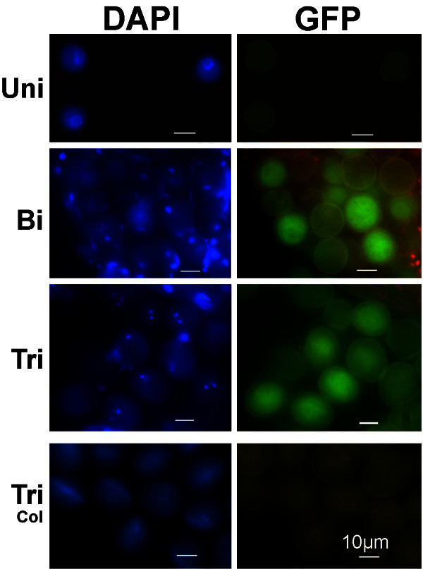
Figure 4.

GFP labelling in pollen grains of thepAt5g17340:UidA:GFPmarker line. GFP signal is clearly detected in bicellular (Bi) and tricellular (Tri) pollen grains of the pAt5g17340:UidA:GFP marker line. At the late uninucleate microspore stage (Uni), the pAt5g17340:UidA:GFP marker line GFP signal is very weak or undetectable. Segregation of the GFP signal (GFP versus non GFP - 1:1 ratio) is detected in the bicellular (Bi) and tricellular (Tri) pollen of a plant hemizygous for the GFP marker gene, indicating a single marker gene insertion in the genome. No GFP signal is detected in Col-0 tricellular pollen grains (Tri-Col). Plates show DAPI stained nuclei (left column) of the microspores and pollen grains shown in the right column. Uni- uninucleate microspore; Bi- bicellular pollen; Tri- tricellular pollen; Tri-Col- wild-type Col-0 tricellular pollen. Marker line plant: G65. Scale bars correspond to 10 μm.
