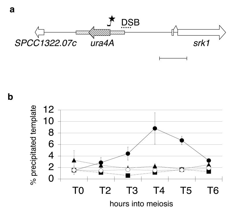
Fig. 1.
Rec12 binds to ura4A hotspot sequences
a. Map of the ura4A insertion used for real time PCR detection of Rec12-binding sequences. ORFs are given as horizontal arrow boxes. The 1.8 kb HinD III ura4 fragment (Grimm et al. 1988), inserted between SPCC1322.07c and srk1, is cross-hatched. Dots indicate the region of DSB formation (Gregan et al. 2005). The location of the ura4A TaqMan® probe is given by a small bar with an asterisk. The length of the scale bar is 1 kb.
b. Graph showing the ratio of real time PCR product from immunoprecipitated chromatin versus total chromatin in rec12myc rad50S (filled circle), rec14Δ rec12myc rad50S (filled triangle), rec6Δ rec12myc rad50S (filled square), and rad50S rec12+(open circle) strains, during meiotic progression in a haploid pat1-114 strain. Independent experiments were performed four times for wild type, three times for rec14Δ, and twice for rec6Δ. Experiments with the untagged control strain were repeated twice. The y-axis gives the ratio of real time template of immunoprecipitated sample to input sample, normalized to 1 μl input, which was set to 100% (see Material and methods). Error bars indicate the standard error of the mean (SEM).

