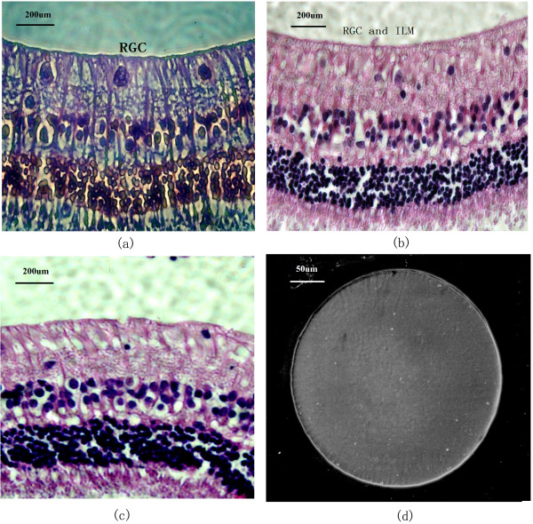
Figure 6.
The morphology of the inner retina layer and microelectrode arrays surface after implantation. (a) Morphology of the inner retinal layer after the microelectrode was implanted for six weeks (retinal ganglion cell layer, RGC; inner limiting membrane, ILM). (phase contrast microscope, 400 ×); (b): rabbit retina layer six months after implant. (light microscope, 400 ×); (c): control retina. (d): SEM of a microelectrode six months after the implant; the electrode showed no damage to the surface or accumulation of tissue matter.

