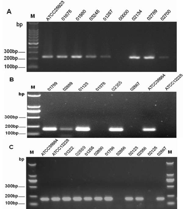
Figure 4.
PCR detection of icaA, icaD, and mecA. Lane 1, 100 bp DNA molecular size marker. Image A shows mecA locus (A, 224-bp band), lanes 2–10, PCR amplification of mecA, lanes 2, ATCC25923. Lane 7, negative control; lanes 3 to 10, PCR amplicons obtained with DNA of S. epidermidis; B shows icaA locus (B, 198-bp band),lanes 2–9, PCR amplification of icaA. Lane 8, ATCC 35984 (RP62A). Lane 9, ATCC 12228. Lanes 2–7, amplification obtained with DNA of clinical S. epidermidis isolate. C shows icaD locus (C, 198-bp band), lanes 2–13, PCR amplification of icaD. Lane 2, ATCC 35984 (RP62A). Lane 3, ATCC 12228. Lanes 4–13, amplification obtained with DNA of clinical S. epidermidis isolate.

