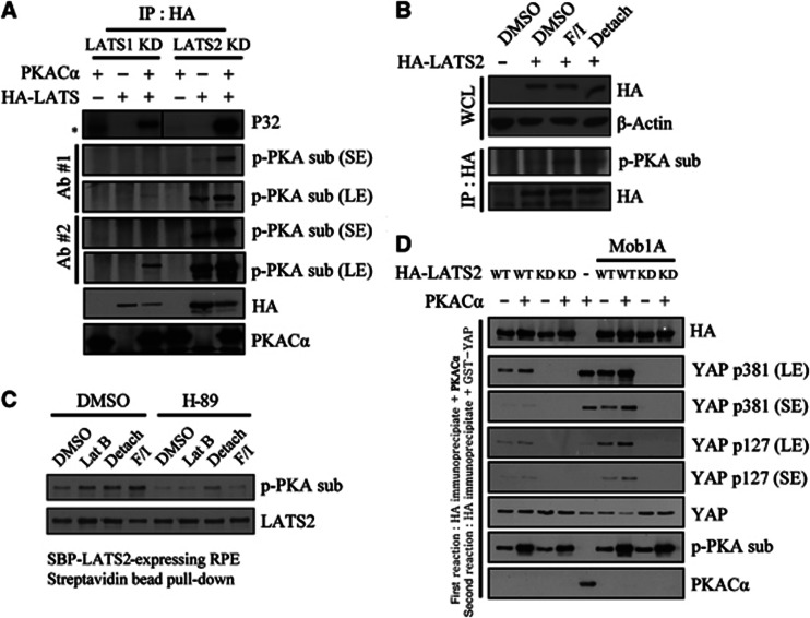
Figure 3.
Phosphorylation of LATS1/2 by PKA enhances LATS kinase activity in cell-free systems and in intact cells. (A) PKA phosphorylates LATS in cell-free system. HA–LATS1 KD (kinase dead) or HA–LATS2 KD was immunoprecipitated from transfected 293T cells. Immunoprecipitated beads were incubated with purified PKACα and radiolabelled ATP. Reaction products were analysed isotopically or by western blotting with phospho-PKA substrate antibodies. *, nonspecific signal. (B) PKA phosphorylates LATS in intact cells. HA–LATS2-transfected NIH3T3 cells were treated with the indicated stimuli for 1 h. After immunoprecipitation with anti-HA antibody, phosphorylation by PKA was examined using a phospho-PKA substrate antibody. WCL, whole cell lysate. (C) RPE cells stably expressing SBP-LATS2 were pre-treated with 20 μM H-89 for 1 h, followed by an additional 1-h treatment with the indicated stimuli. LATS2 was pulled down using Streptavidin agarose bead and assayed as in panel (B). (D) LATS2 pre-incubated with PKA has increased kinase activity. HA–LATS2 WT or KD mutant was immunoprecipitated from 293T cells and reacted with PKACα. PKACα was extensively washed out, followed by incubation with 1 μg GST–YAP (full-length) protein and 200 μM unlabelled ATP. Reaction products were analysed by SDS–PAGE and immunoblotting. In lanes 6–9, HA–Mob1A was co-transfected to increase overall kinase activity. In lane 5, GST–YAP was reacted with PKACα to examine possible background phosphorylation of YAP by PKA. SE, short exposure; LE, long exposure.

