
Figure 4.
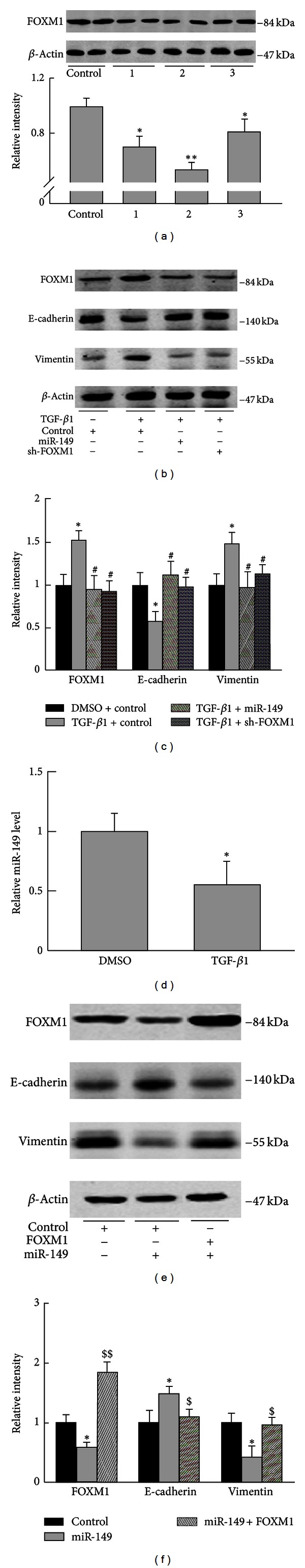
FOXM1 was involved in EMT of A549 cells induced by TGF-β1. (a) A549 cells were transfected with sh-control, sh-FOXM1 1~3, respectively, for 48 h and harvested for western blot. sh-FOXM1 2 showed the most inhibitory effect and was selected for the following experiments. (b) A549 cells were transfected with control, sh-FOXM1, or miR-149, respectively, and incubated for 24 h, then TGF-β1 (5 ng/mL) was added to the medium, and cells were harvested 48 h later. Western blot examined the protein levels of FOXM1, E-cadherin, and vimentin. (c) Relative intensity of blots in (b) was analyzed. (d) A549 cells were treated with DMSO or TGF-β1 (5 ng/mL) for 24 h and collected for microRNA extraction. miR-134 expression was examined. (e) A549 cells were transfected with control, miR-149, or cotransfection of miR-149 and FOXM1 for 48 h. Cells were harvested for western blot analysis. (f) Relative intensity of blots in (e) was analyzed. Data were from three independent experiments. *P < 0.05, compared with control group; # P < 0.05, ## P < 0.01, compared with TGF-β1 group; $ P < 0.05, $$ P < 0.01 compared with miR-149 group.