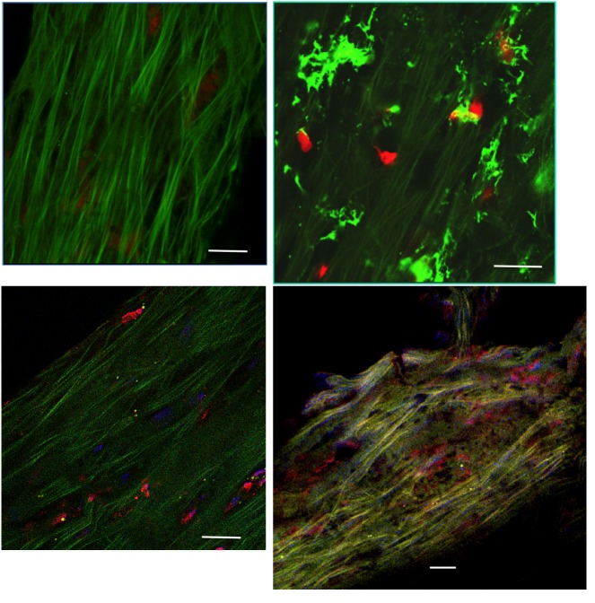
FIGURE 1.
Immunohistological assessment of human trabecular meshwork (TM) specimens examined by confocal microscopy. Top left, A few TM cells, visible through their red nuclei, scattered throughout a dense green autofluorescent matrix. Top right, Dendritiform inflammatory cells infiltrating the TM (vimentin green staining). Bottom left, Trabecular cells expressing fractalkine (red immunostaining). Bottom right, Trabecular cells expressing CX3CR1 (red immunostaining). (Red nuclear staining by propidium iodide (top), blue staining by DAPI (bottom). Bars = 50 μm.

