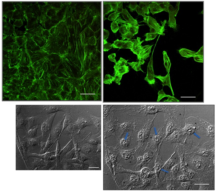
FIGURE 2.
Cytological assessment of proapoptotic effects of benzalkonium (BAK) in trabecular meshwork (TM) cells. Top, Phalloidin staining shows actin cytoskeleton arrangement in HTM3 cells submitted to 0.01% BAK for 15 minutes. Control cells are at left. At right, BAK-stimulated cells show major shrinkage and apoptotic features. Bottom, Phase contrast examination of TM cells submitted to BAK 0.01%. Normal state at baseline at left. At right, after 5 minutes of contact, retraction and fragmentation of nuclei can be observed (arrows) with leakage of cytoplasmic material in response to membrane disruption (stars). Bars = 50 μm.

