
FIGURE 6.
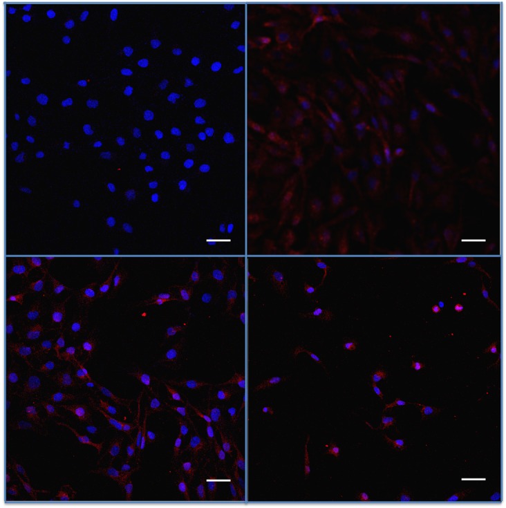
Assessment of fractalkine receptor CX3CR1 in trabecular meshwork cells submitted to benzalkonium (BAK) or TNF-α. Indirect immunostaining for CX3CR1 (in red) in trabecular cells (nuclei in blue). Top left, Negative fluorescence intensity in basal conditions. Top right, Increased expression after TNF-α. Bottom left, BAK 5×10⁻⁵%. Bottom right, BAK 5×10⁻⁴% stimulations for 24 hours. Bars = 50 μm.