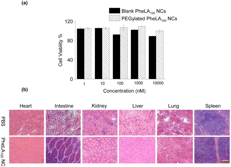
Figure 6.
(a) Cytotoxicity of blank PheLA100 NCs and PEGylated PheLA100 NCs in MCF-7 cells over 72 h at 37°C, determined by MTT assays. (b) Histopathology analysis of mouse tissues following an i.v. injection of PEGylated PheLA100 NCs via tail vein. Representative sections of various organs taken from the control mice receiving PBS and the treatment mice receiving 250 mg/kg PEGylated PheLA100 NCs 24 h post injection were stained by hematoxylin and eosin. No organs of the mice given PEGylated PheLA100 NCs showed acute inflammations.

