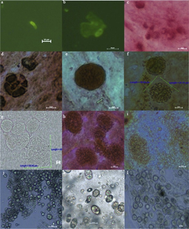
Fig. 2.
Sporozoites, trophozoites, schizonts, gamonts and oocysts in embryos at various times after inoculation (a-l). (a) 2 h p.i., sporozoites; (b) 12 h p.i., sporozoites in the CAM cell; (c) 24 h p.i., trophozoites; (d) 48-h p.i., immature schizonts; (e) 60 h p.i., mature first-generation schizonts; (f) 72 h p.i., mature first-generation schizonts and merozoites; (g) 96 h p.i., second-generation schizonts and merozoites; (h) 120 h p.i., second-generation schizonts and merozoites; (i) 144 h p.i., immature macrogametes and microgametocytes; (j) 168 h p.i., mature macrogametes and microgametocytes; (k) 192 h p.i., oocysts and immature oocysts; (l) nine-days p.i., mature unsporulated oocysts.

