
Figure 3.
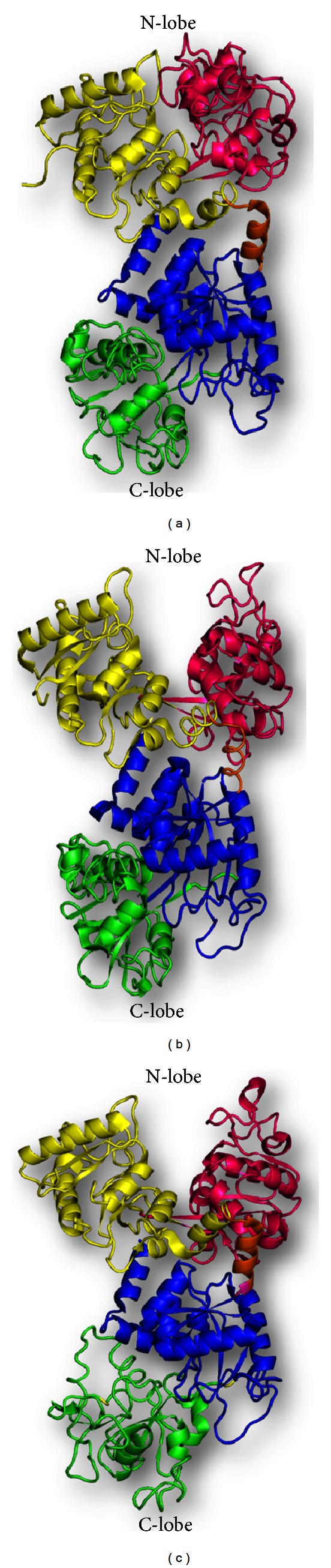
Schematic diagrams of apolactoferrins from three species showing variable behavior of the conformation of the domains. (a) Equine apolactoferrin (PDB code: 1B7U) shows both the lobes in closed conformation. (b) Human apolactoferrin (PDB code: 1CB6) shows open N-lobe and closed C-lobe. (c) Camel apolactoferrin (PDB code: 1DTZ) shows both the lobes in open conformation.