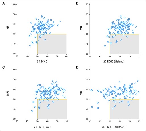
Fig 3.
Scatter plots identifying survivors with an ejection fraction by cardiac magnetic resonance imaging (MRI) of less than 50% but ≥ 50% (gray box) by echocardiography (ECHO) for (A) three-dimensional (3D) echocardiography, (B) two-dimensional (2D) biplane, (C) 2D apical 4-chamber (A4C), and (D) 2D Teichholz method.

