
Figure 3.
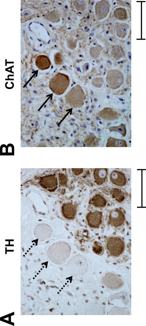
TH-negative cells are ChAT-positive in the LSG A, Representative TH-staining of the LSG from a Group 1 dog. Dashed arrows point to three TH-negative ganglion cells. B, A serial slide from the same LSG is used for ChAT immunostaining. The same three ganglion cells that are TH-negative are positive for ChAT (arrows). This development of cholinergic phenotype is observed in a vast majority of TH-negative cells, whose number is increased by LL-VNS. TH, tyrosine hydroxylase; ChAT, choline acetyltransferase. Calibration bar = 50 μm.