
Figure 4.
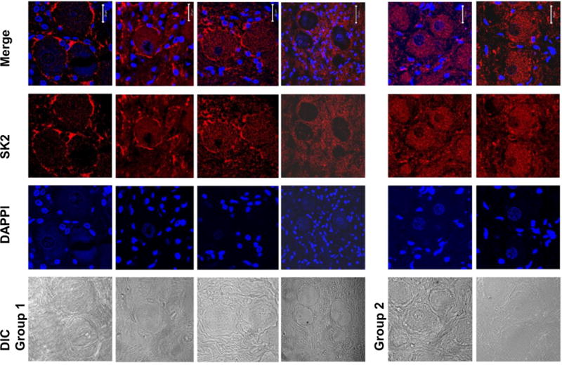
LL-VNS changes the subcellular distribution of SK2 protein. Immunofluorescence confocal microscope images of four Group 1 (LL-VNS) LSG and two Group 2 (control) LSG using anti-SK2 antibody and DAPI (4′,6-diamidino-2-phenylindole to identify nuclei). Corresponding differential interference contrast (DIC) images are shown on the left. The merged picture shows that in Group 1 LSG, there was a significant increase in SK2 staining in the periphery of ganglion cells but decrease in the cytosol. In contrast, in Group 2 LSG, the SK2 staining was homogeneous. DAPI, 4′, 6-diamidino-2-phenylindole. The white calibration bars are 20 μM in length.