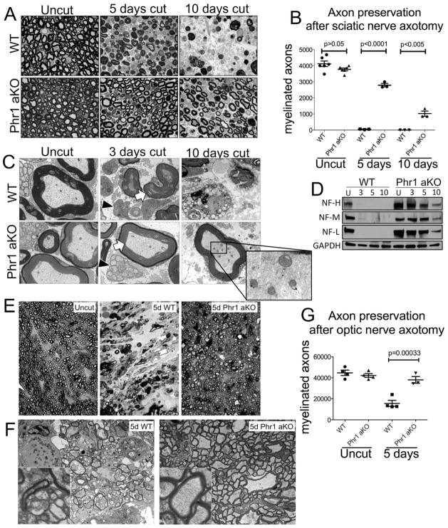
Figure 1.
Phr1 adult conditional KO (Phr1 aKO) delays axon degeneration in the CNS and PNS. (a) Toluidine-blue stained cross-sections of axotomized sciatic nerve distal to the cut site, showing delayed axon degeneration in Phr1 aKO compared to littermate controls (WT). (b) Quantification of intact myelinated axons of cut and unlesioned contralateral sciatic nerves. (c) Electron micrographs of the nerves shown in (a). Black arrowheads point to preserved small unmyelinated sensory fibers (Remak bundles) in Phr1 aKO nerves. White arrows point to the preservation of large caliber axons, whose cytoskeleton and organelles remain intact for at least 10 days (inset). (d) Representative Western blot of tibial nerves at the indicated time points after transection, showing preservation of neurofilaments (heavy, NF-H; medium, NF-M; light, NF-L chains) in Phr1 aKO mice (n=3). (e) Toluidine-blue stained cross-sections of optic nerve distal to the cut site demonstrate almost complete preservation of axons in Phr1 aKO mice five days after eye enucleation, a time-point when WT optic nerves are extensively degenerated, as quantified in (g). (f) Electron micrographs of the same optic nerves shown in (e), demonstrating axon death in WT samples, and morphological axon preservation in Phr1 aKO samples (insets) five days after lesion.

