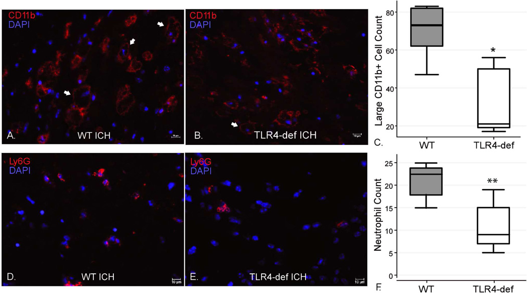
Figure 1. Immunohistochemical staining for neutrophils and myeloid-derived cells 3 days after ICH surgeries in WT and TLR4-deficient mice.
(A–B) There was no significant difference between the genotypes in total numbers of CD11b+ cells in five 20x fields (WT 255.6 ± 77.3 cells vs. TLR4-deficient 218.4 ± 70.1 cells, p=0.45). However, there were more large, vacuolated CD11b+ cells in the WT mice (representative cells indicated by arrows). (C) Boxplot of the number of large (>10 µm), vacuolated CD11b+ cells in five 20x fields per mouse by genotype. * p<0.05, n=5/genotype. (D–E) Ly6G staining for neutrophils demonstrated more perihematomal neutrophils in the WT mice compared to TLR4-deficient mice. (F) Boxplot of numbers of perihematomal neutrophils in five 40x fields per mouse by genotype. ** p<0.01, n=5/genotype. scale bar indicates 10 µm.

