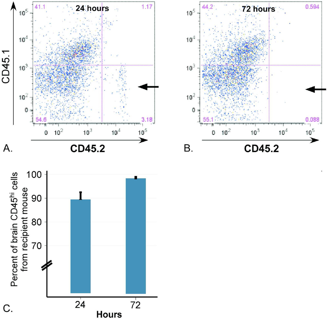
Figure 3. Source of perihematomal leukocytes on post-ICH days 1 and 3.
Injection of blood from C57BL/6 mice (homozygous for the pan-leukocyte marker CD45.2) into B6LY5.2/Cr mice (homozygous for CD45.1) revealed that the majority of leukocytes quantified by flow cytometry originate from the recipient mouse. (A) Representative plot demonstrating only 3.2% of live cells staining positive for CD45.2 (arrow) at day 1 post-ICH, indicating their origin from the injected ICH. (B) At day 3 post-ICH, less than 0.1% of live cells are CD45.2+ (arrow). (C) Quantification of percent of ipsilateral CD45+ cells deriving from the recipient mouse (CD45.1+) at each time point.

