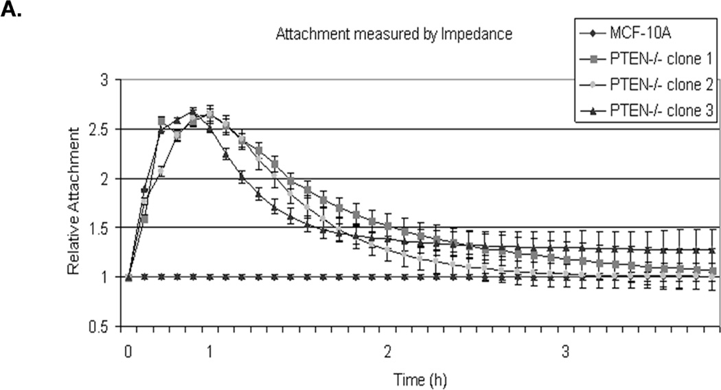
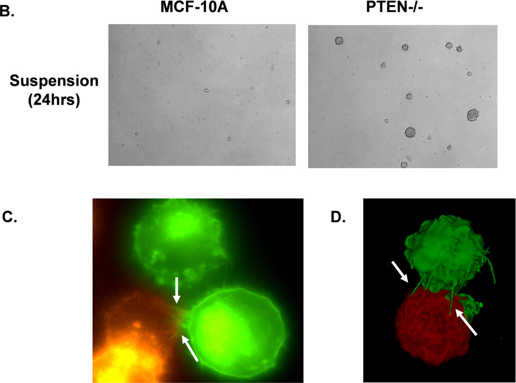
Figure 2. Attachment and homotypic aggregation are enhanced in PTEN−/− cells.


A. A greater change in impedance of the PTEN−/− cells illustrates an increase in attachment compared to the MCF-10A cells. Impedance values are normalized to MCF-10A (n=4, representative results from triplicate experiments). B. An equal number of cells were suspended in normal growth media in ultra-low attachment plates. After 48h, only the PTEN−/− cells aggregate into tight, spheroid structures. C. Live cell imaging of two cell populations of the same PTEN clone, one GFP-mem transfected (green) and the other CellMask stained (red), were combined and suspended over a BSA coated glass surface to prevent attachment. Arrows indicate McTNs extending from the GFP-mem cell attaching to a CellMask stained cell. D. 3D computer rendered image of PTEN−/− cells from separate populations (GFP-mem transfected and CellMask stained) associating. Arrows indicate McTNs from the GFP-mem cell contacting the CellMask stained cell.
