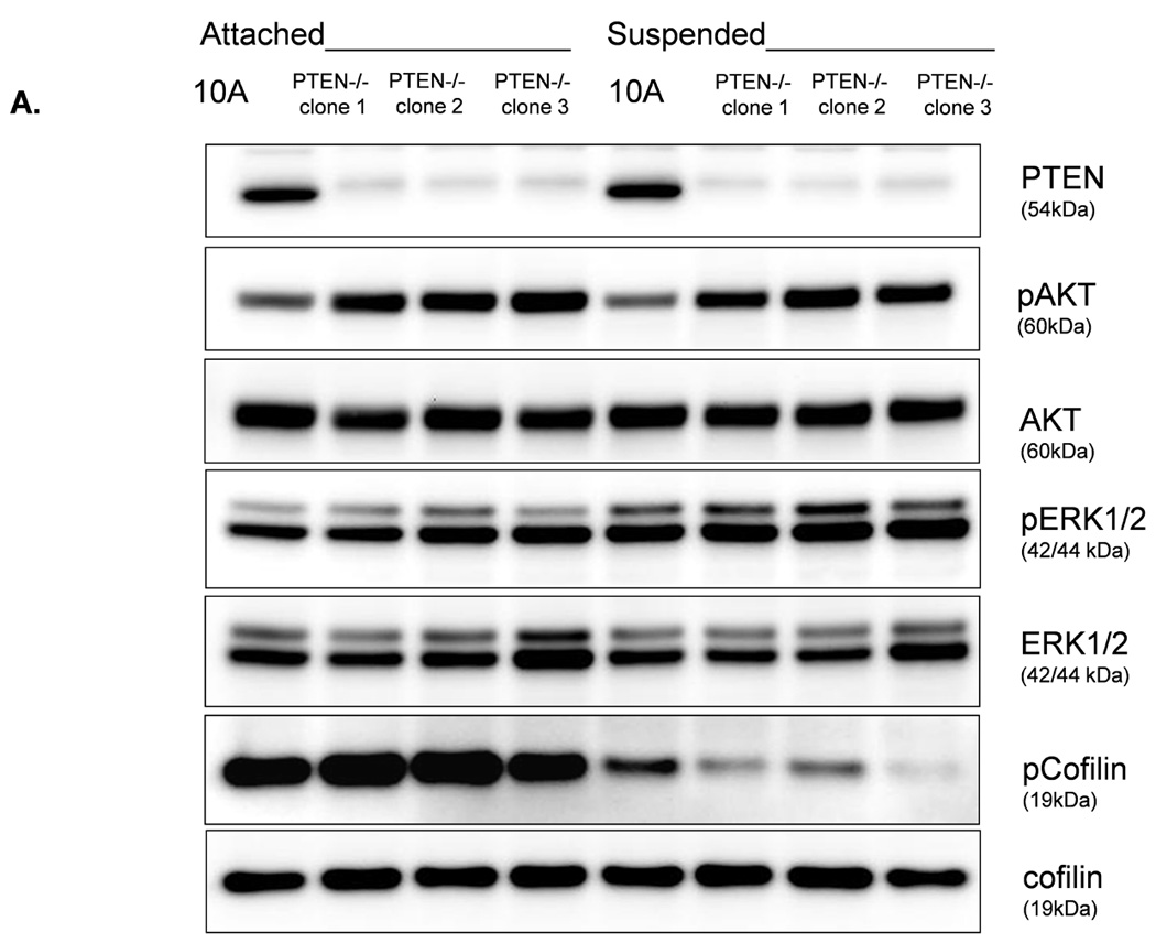
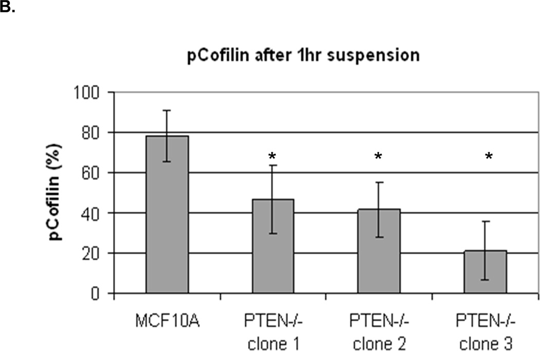
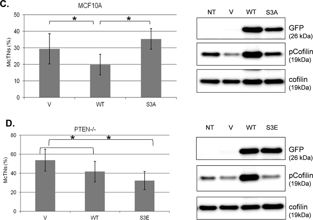
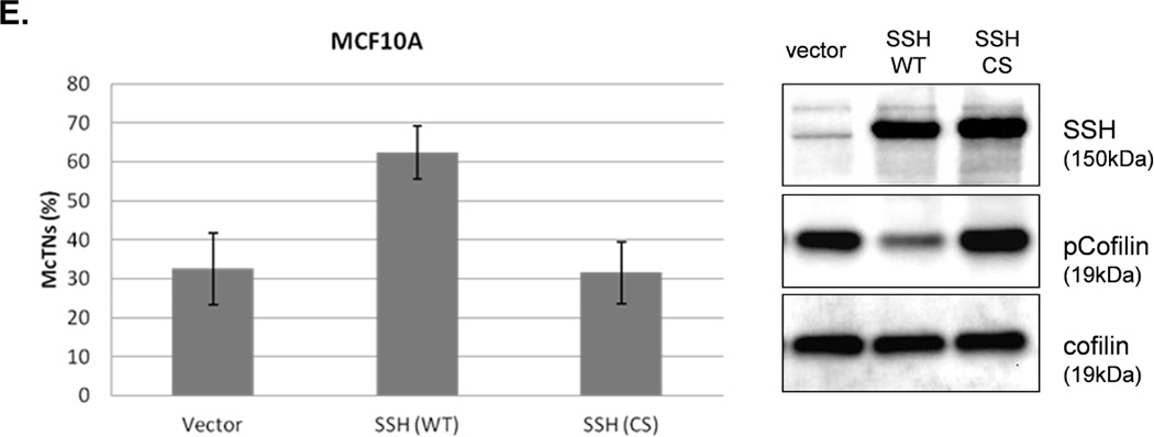
Figure 4. Cofilin is activated in suspended cells, but to a much higher degree in cells without PTEN expression.


A. Representative Western blot analysis shows that cofilin is highly phosphorylated (pCofilin) and inactive when cells are attached and dephosphorylated to an active form when cells are detached. B. Densitometry analysis shows that pCofilin levels only decrease 20% in MCF-10A cells after detachment. By comparison, pCofilin levels are reduced 55–75% in PTEN−/− cells, indicating a more robust detachment-induced activation of cofilin in PTEN−/− cells. Levels of pCofilin were normalized to total cofilin in each sample and the ratio of pCofilin in suspended cells compared to attached cells was determined (n=4, *p<0.01). C. MCF-10A cells transfected with vector only (V), cofilin (WT), or mutant cofilin (S3A) and D. PTEN−/−cells transfected with vector only (V), cofilin (WT), or mutant cofilin (S3E) for 24h. Untransfected (NT) and transfected cells were blindly counted for McTNs (n=6, *p<0.01) and analyzed by Western blotting. E. MCF-10A cells were transfected with vector only (V), wild-type Slingshot-1L (SSH WT), or catalytically inactive Slingshot-1L (SSH CS) for 24hr and scored blindly for McTNs (n=6, *p<0.01) or analyzed by Western blotting.
