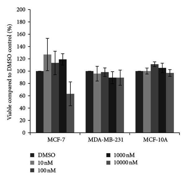
Figure 6.

Cytotoxicity of chlorpyrifos. MCF-7 and MDA-MB-231 mammary epithelial carcinoma and MCF-10A mammary epithelial cells were plated in a 96-well plate at 6000 cells per well and treated with 10 nM to 10000 nM chlorpyrifos or DMSO vehicle control for 48 hours. Live cells were stained with crystal violet and the amount of dye absorbed was quantified by a microplate reader at 450 nm. Results are shown as percentage of live cells compared to DMSO control set at 100 percent. Significant variation and trends of variation from the control were calculated by student's t-test (a*, P < 0.05).
