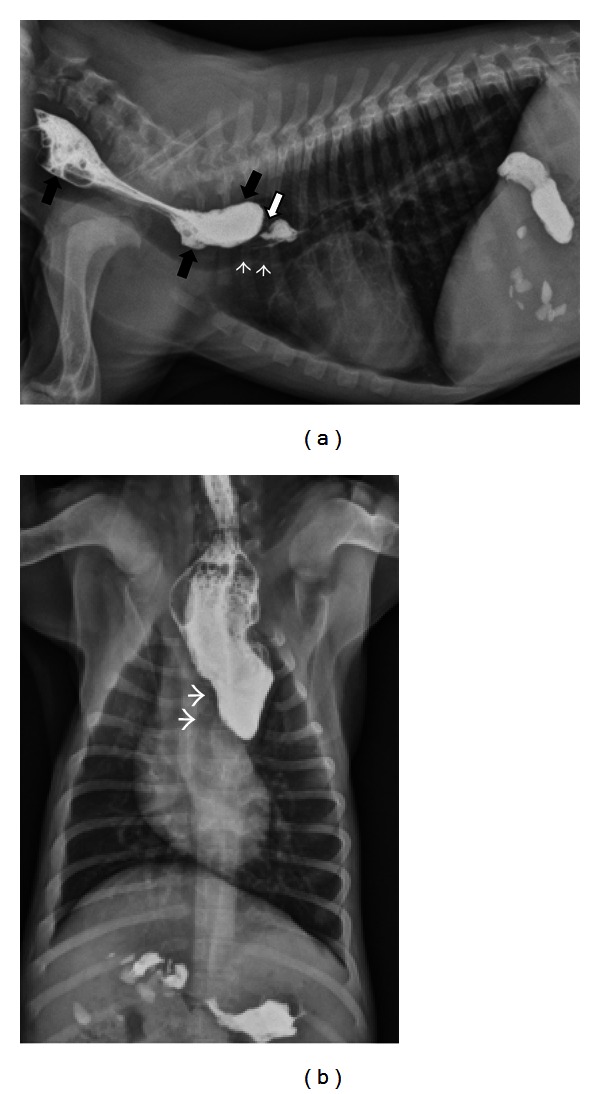
Figure 3.

(a) A right lateral thoracic radiograph depicts a 2-month-old male Labrador presenting for regurgitation starting after weaning. Liquid barium has been administered orally immediately prior to radiography. There is a focal narrowing of the esophageal luminal diameter immediately dorsal to the heart base (white arrow). The trachea undulates and deviates ventrally in the same region (white arrowheads). Contrast is pooling is a dilated esophagus oral to the lesion (black arrows). (b) A dorsoventral radiograph of the same dog as in (a) shows contrast dilation of the cervical esophagus and an abrupt termination of the contrast column at the heart base. The trachea deviates to the left (white arrowheads) as is typically seen with persistent right aortic arch.
