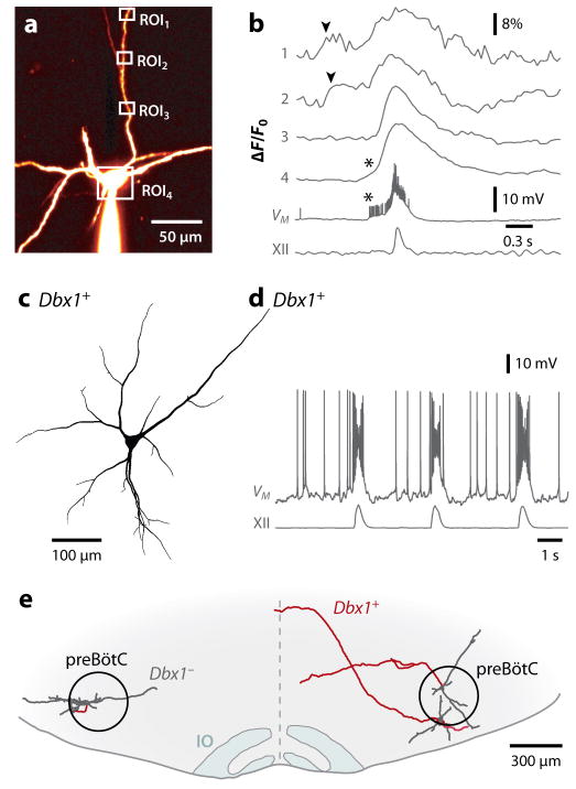
Figure 3. Inspiratory burst generation: role of dendrites and properties of Dbx1+ preBötC neurons.
A and B, dendritic two-photon Ca2+ imaging and somatic patch clamp. A, preBötC neuron filled with fluorescent dye from somatic whole-cell recording. Regions of interest (ROIs) shown, which correspond to B. B, dendritic Ca2+ transients (arrowheads) precede somatic bursts and XII motor output. Asterisks indicate somatic spike-driven Ca2+ transients (Del Negro et al 2011). C and D, morphology (C) and physiology (D) of a Dbx1+ preBötC neuron. Drive potentials of ~25 mV amplitude and depolarization block of spiking indicative of ICAN activation during the inspiratory burst. E, transverse view of a mouse slice (ventral) showing two Dbx1+ neurons (right) and a Dbx1− neuron (left) recorded and biocytin reconstructed. All three were inspiratory neurons. Dbx1+ neurons are commissural (axons in red). IO is inferior olive.

