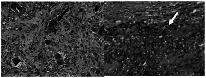
Figure 4.

An open biopsy was performed. Microscopic examination revealed a nodular growth covered by synovial lining cells. Mitotic figures and a large number of multinucleated giant cells (black arrow, left) were observed in certain areas. Inflammatory cells were identified in various areas and the stroma demonstrated fibrosis. The white arrow indicates hemosiderin deposition (right). Histological findings were characteristic of pigmented villonodular synovitis.
