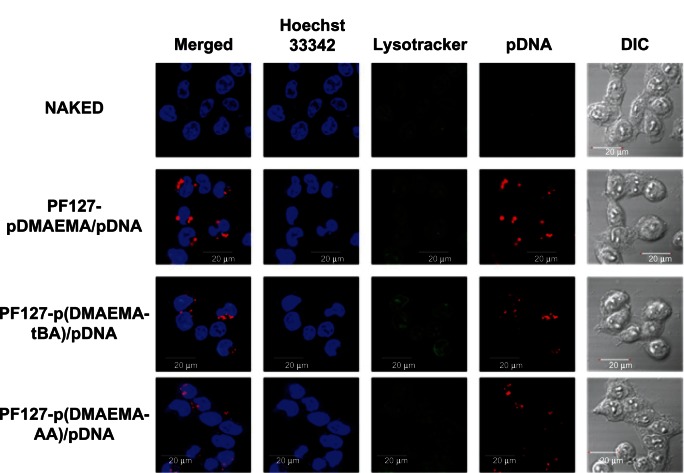
Figure 9.
CLSM images of 293T cells exposed to copolymer/Cy5-labeled pGL3 polyplexes at N/P = 9 for 4 hours of incubation.
Note: Blue: nuclei (Hoechst 33342), green: endolysosome (Lysotracker), and red: Cy5-labeled pGL3 plasmid DNA.
Abbreviations: CLSM, confocal laser scanning microscope; N/P, nitrogen/phosphate; pDNA, plasmid deoxyribonucleic acid; DIC, differential interference contrast; PF127-pDMAEMA, pluronic F127-poly (dimethylaminoethyl methacrylate); PF127-p(DMAEMA-tBA), pluronic F127-poly (dimethylaminoethyl methacrylate-tert-butyl acrylate); PF127-p(DMAEMA-AA), pluronic F127-poly (dimethylaminoethyl methacrylate-acrylic acid).

