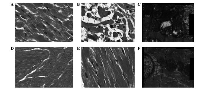
Figure 1.
(A) Adriamycin-induced myocardial injuries and muscle bundle fractures were visible under a light microscope with low magnification in the ADM group (magnification, ×100); (B) Adriamycin-induced myocardial mucinous degeneration was visible under a light microscope with high magnification in the ADM group (magnification, ×400); (C) Adriamycin-induced organelle injuries in myocardial cells and serious mitochondrial cavitation were visible under a light microscope with high magnification in the ADM group (magnification, ×20.0KX); (D) Essentially normal cardiac muscles were visible under a light microscope with low magnification in the M+A group (magnification, ×100); (E) Only mild granular changes were visible under a light microscope with high magnification in the M+A group (magnification, 400×); (F) Adriamycin-induced organelle injuries in myocardial cells and serious mitochondrial cavitation were visible under an electron microscope with high magnification in the M+A group (magnification, ×20,000). Blank, no intervention; Diss, solvent intervention; MLT, melatonin intervention; ADM, Adriamycin intervention; M+A, melatonin + Adriamycin intervention.

