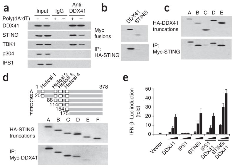
Figure 6.
Interaction of DDX41 with STING. (a) Immunoblot analysis of proteins (left margin) precipitated with anti-DDX41 or immunoglobulin G (IgG; control) from whole-cell lysates of D2SC cells left unstimulated (−) or stimulated with poly(dA:dT) (+). Input, 10% of the D2SC cells lysate. (b) Immunoblot analysis of immunoprecipitation assays of purified HA-tagged STING incubated with Myc-tagged DDX41 or STING, probed with anti-Myc. (c) Immunoblot analysis of immunoprecipitation assays of purified HA-tagged full-length or truncated DDX41 (as in Fig. 4b) incubated with Myc-tagged STING, probed with anti-HA. (d) Immunoblot analysis of purified HA-tagged full-length STING (A) or truncated STING (B–F), probed with anti-HA (middle), and immunoblot analysis of immunoprecipitation assays of HA-tagged STING (as above) incubated with Myc-tagged DDX41, probed with anti-HA (bottom). Top, full-length STING (A) and serial truncations of STING (B–F); numbers indicate positions of amino acids. (e) Activation of the Ifnb promoter in mouse L929 cells transfected with an IFN-β luciferase reporter (IFN-β–Luc; 100 ng) plus increasing concentrations (20, 100 or 200 ng; wedges) of expression vectors for DDX41, IPS1 or STING individually; or expression vector for DDX41 (20 ng; solid bar) together with increasing concentrations (20, 100 or 200 ng; wedges) of expression vectors for IPS1 or STING. Results are presented relative to those of cells transfected with empty vector alone (Vector). Data are representative of three independent experiments (mean and s.d. in e).

