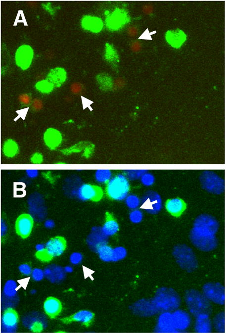
Fig. 2.
Dcx+ cells (green) demonstrate signs of apoptotic cell death (red staining with Magic Red Live caspase 3&7 reagent), arrows, after 12 h of treatment with the mitochondrial inhibitor antimycin A (2 μM) (A). Note rapid disappearance of green Dcx staining in apoptotic cells. The bottom panel shows Dcx and nuclear DAPI staining of the same area

