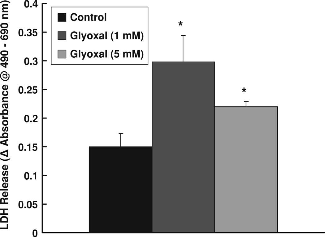
Fig. 4.
Glyoxal induced LDH release by endothelial cells. BPAECs (90% confluent), 17.5-mm dishes (24-well culture plate) were treated with MEM alone or MEM-containing glyoxal (1 and 5 mM) for 12 h at 37°C in a humidified environment of 5% CO2–95% air. At the end of incubation, release of LDH into the medium (as an index of cytotoxicity) was determined spectrophotometrically as described under Materials and methods section. Data represent means ± SD of three independent experiments. * Significantly different at P < 0.05 as compared to the cells treated with MEM alone

