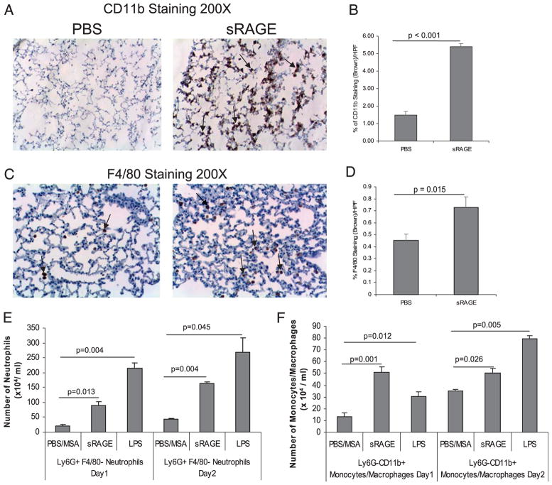
FIGURE 1.
sRAGE induces macrophage and neutrophil infiltration in the murine lung in vivo. C57/BL6 mice were injected IT with 5.0 μg sRAGE per gram of mouse weight, 200 μg LPS, or 200 μg PBS/MSA for 1 or 2 d. Lungs were harvested and subjected to immunohistochemical staining for CD11b (A) or F4/80 (C) expression. Ten pictures per slide were taken using an Olympus 1 × 50 inverted microscope equipped with a 20× objective and Nikon camera (Olympus). Brown staining represents CD11b+ cells (A) or F4/80+ cells (C). The images were quantified by histogram analysis using Adobe Photoshop CS (B, D). Shown are representative pictures obtained from three pairs of mice. Single-cell suspension from the lungs were also stained with Ly6G, F4/80, and CD11b Abs and analyzed by flow cytometry. Neutrophils were defined as Ly6G+ F4/80− population (E), and monocytic cells were defined as Ly6G–CD11b+ population (F). Data presented are the percent positive cells from the total cell number obtained from at least three different mice and are expressed as the mean ± SEM.

