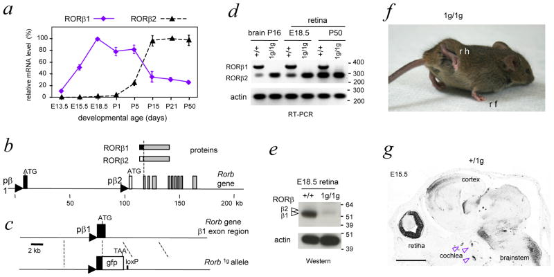
Figure 1. Differential expression of RORβ isoforms and targeted deletion of RORβ.
1
a, RORβ1 and RORβ2 mRNA expression profiles in mouse retinal development, determined by qPCR analysis. Levels for each isoform are indicated as percentages relative to the highest level, assigned a value of 100%.
b, Rorb gene with RORβ1- and RORβ2-specific exons depicted as black and white boxes, respectively, common exons as grey boxes and RORβ1 and RORβ2 promoter regions as triangles.
c, Targeted replacement of the coding portion of the RORβ1-specific exon with a green fluorescent protein (gfp) cassette that carries a TAA translational stop codon. Excision of the neomycin-resistance ACN cassette left a residual loxP site in the Rorb1g allele.
d, Loss of RORβ1 mRNA and retention of RORβ2 mRNA in brain and retina in 1g/1g mice shown by PCR with control analysis for β-actin. DNA size marker, bp.
e, Loss of RORβ1 protein in the retina in 1g/1g mice shown on a western blot with control analysis for β-actin. Arrowheads, bands for RORβ1 (lower, ~52 kDa) and RORβ2 (upper, ~53 kDa). The gfp protein expressed in 1g/1g mice carries no RORβ protein sequence and was detected with antibody against gfp as a ~27 kDa band (not shown). Protein molecular size marker, kDa.
f, Abnormal gait in 1g/1g mice with exaggerated lifting and clasping of hindlimbs; rh, right hindlimb; rf, right forelimb.
g, Para-sagittal section of +/1g embryonic head showing gfp expression in retina, cochlea and brain. Expression of gfp was detected by immunohistochemistry (dark areas). Scale bar, 1mm.

