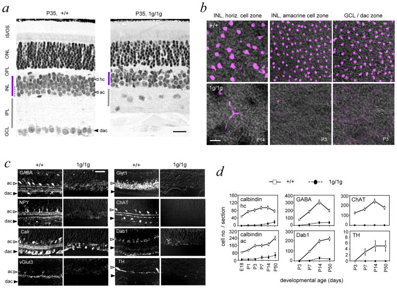
Figure 3. Loss of amacrine and horizontal cells in Rorb1g/1g mice.
a, Histological sections showing disorganized inner plexiform, outer plexiform and ganglion cell layers in 1g/1g mice at P35. Note the thin INL (purple bar) and mis-located ganglion cell soma in the collapsed IPL in 1g/1g mice. Amacrine cell zone in the INL is indicated by a dashed line in +/+ retina. Scale bars, 30 μm in all panels.
b, Retinal flatmounts analyzed by confocal microscopy in the plane of horizontal (hc), amacrine (ac) and displaced amacrine (dac) cell zones using calbindin as a marker (purple) on DIC images to show tissue structure. Calbindin+ horizontal and amacrine cells are missing in 1g/1g mice.
c, Retinal sections showing loss of representative amacrine cell markers (GABA, GlyT1, NPY, ChAT, calretinin/calr, Dab1, vGlut3, TH) in 1g/1g mice at P14 (except NPY at P7).
d, Cell counts showing failure to generate amacrine and horizontal cells, identified with horizontal (calbindin/hc) and amacrine (calbindin/ac, GABA, ChAT, Dab1, TH) markers. Calbindin+ horizontal and amacrine cells were distinguished in +/+ mice by morphology and location in the INL. Cell counts, given as mean ± S.D., were based on analysis of 12 cryosections of the retina representing ≥ 3 mice at each age.

