
Fig. 3.
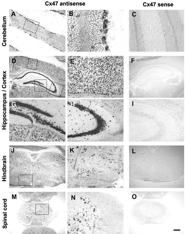
Expression of Cx47 mRNA in the murine CNS. Coronal sections of adult brain and spinal cord were hybridized as described in Materials and Methods. Representative sections from the cerebellum (A–C), forebrain (D–I), hindbrain (J–L), and spinal cord (M–O) are shown. Note that the in situ signal labels preferentially and/or cell layers known to contain specific neuronal phenotypes, such as the Purkinje cell layer and the granule cell layers in the cerebellum (A, B). Within the hippocampal formation (D, G, H), heavy labeling is seen over the dentate gyrus (G) and all subregions of the cornu Ammonis (D, H; the latter micrograph depicts the CA3 region). Within the cerebral cortex (D, E), numerous cells located in all cell layers are positive. In the brainstem, labeling was particularly prominent over nuclei, such as the inferior olive (J, K). The micrographs from the cervical spinal cord (M, N) again show numerous labeled cells within the gray matter and also document that oligodendrocytes and astrocytes within the white matter do not express detectable levels of Cx47 mRNA. Sections shown inA, B, D, E, G, H, J, K, M, and N were hybridized with the Cx47 antisense probe. Controls shown in C, F, I, L, and O were hybridized with a Cx47 sense probe. Rectangles in micrographs A, D, J, and M indicate the areas from which the higher power views shown in B, E, G, H, K, and N, respectively, were taken. Scale bar: A, C, E, G–I, K, N, 100 μm; B, 25 μm; D, F, J, L, M, O, 400 μm.