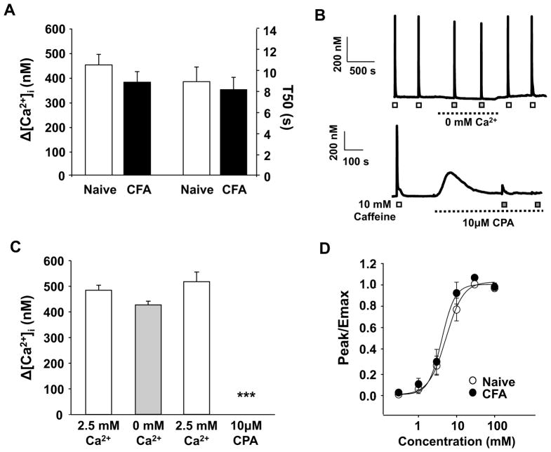
Figure 3.
Inflammation did not change in the magnitude or duration of the caffeine-evoked Ca2+ transient. A) The magnitude (Δ [Ca2+]i) and duration (T50) of caffeine (10 mM for 4 sec) evoked Ca2+ transients were determined in a manner identical to that used for the high K+-evoked transient. Pooled data from putative nociceptive cutaneous neurons from naïve (n = 36) and inflamed (n = 29) rats are plotted. B) Top: Example of caffeine-evoked Ca2+ transients in a putative nociceptive cutaneous neuron from a naïve rat in the presence (open square) and absence (closed square) of Ca2+ in the bath solution. Bottom: Example of caffeine-evoked Ca2+ transients in a putative nociceptive cutaneous neuron from a naïve rat before (open square) and after (closed square) the application of the SERCA inhibitor, CPA. C) Pooled data from neurons (n = 8) challenged with caffeine before and after the application Ca2+ free bath solution and from a second groups of neurons (n = 10) challenged before and after depletion of intracellular Ca2+ stores with CPA. D) Pooled concentration response data for putative cutaneous neurons from naïve (n = 13) and inflamed (n = 14) rats challenged with increasing concentrations of caffeine. Data from each neuron were fitted with a modified Hill equation to estimate the maximal size of the evoked transient (Emax) and this value was used to normalize data prior to pooling. Pooled data are also fitted with a modified Hill equation. ***p < 0.001

