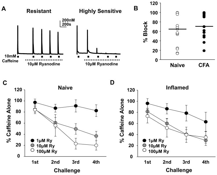
Figure 5.
Heterogeneity in the ryanodine receptor activity in neurons from both naïve and inflamed rats. A) Examples of caffeine-evoked Ca2+ transients in putative nociceptive cutaneous neurons before and after the application of 10 μM ryanodine in Ca2+ free bath. The neuron on the left was relatively resistant to ryanodine-induced block, while that on the right was highly sensitive. B) The percent of ryanodine-induced block of the caffeine-evoked Ca2+ transient was calculated from the response evoked by the 4th caffeine application relative to the response evoked prior to the application of ryanodine. Data from neurons from naïve (n = 15) and inflamed (n = 15) rats challenged with 10 μM ryanodine are plotted. C) Neurons (n = 31) from naïve rats were challenged with caffeine (10 mM) before and after the application of 1 of 3 concentrations of ryanodine (Ry). The response to each of 4 successive applications of caffeine was calculated as a percent of the response to caffeine prior to the application of caffeine. Pooled data over subsequent challenges are plotted relative to the concentration of ryanodine employed. D) Data from neurons (n = 33) from inflamed rats were collected and plotted as described in C. There was no significant interaction between inflammation and ryanodine concentration despite significant main effects associated with caffeine application and ryanodine concentration (Mixed 3 way ANOVA, p > 0.05).

