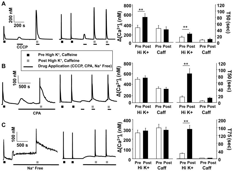
Figure 8.
High K+- and caffeine-evoked Ca2+ transients engage distinct Ca2+ regulatory machinery. Putative nociceptive cutaneous DRG neurons from naive rats were challenged with either high K+ (30 mM, 4s, Left Traces) or caffeine (10 mM, 4s, Right Traces), before and after the application of CCCP (10 μM, A), CPA (10 μM, B), or Na+ Free bath (C). Pooled magnitude (peak change in concentration of intracellular Ca2+ ([Ca2+]i) from baseline) and duration (time of decay) data obtained before and after the application of CCCP, CPA and Na+ Free bath are plotted. For the high K+ experiments, the number of neurons studied were 7, 11, and 12, with CCCP, CPA and Na+ free bath, respectively, while for the caffeine experiments, the number of neurons studied were 6, 6 and 6 for CCP, CPA and Na+ free bath, respectively. Data were analyzed with a mixed design two-way ANOVA: differences between before and after the application of CCCP, CPA or Na+ Free bath, as well as between high K+ and caffeine are indicated. * is p < 0.05, and ** is p < 0.01.

