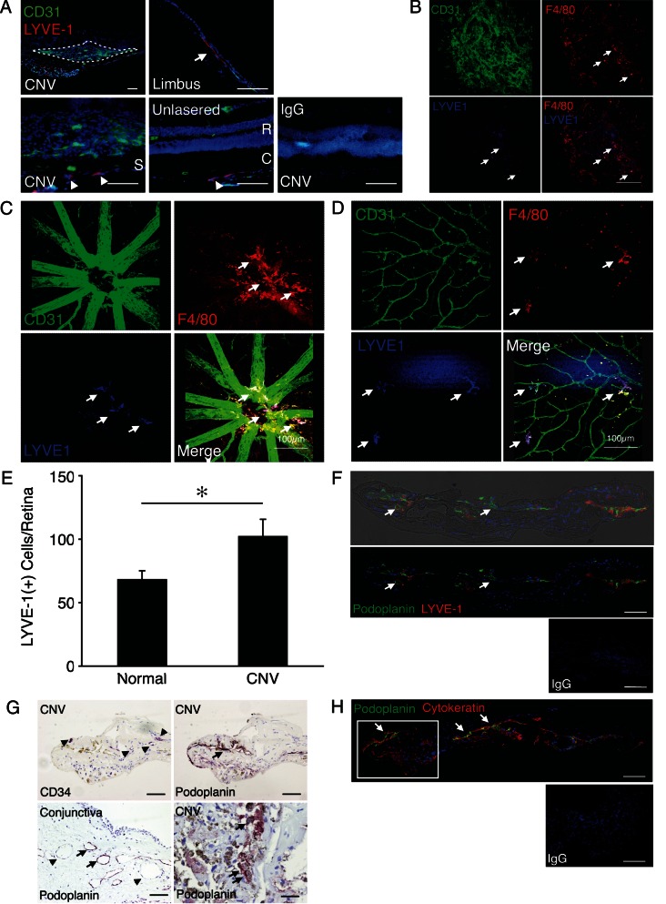
Figure 1.
Lymphatic markers in the mouse CNV model and in human CNV membranes. (A) Double immunostaining of laser-induced CNV (day 7) (left), corneal limbus (top right), and normal retina (R) and choroid (C) with antibodies against CD31 (green), LYVE-1 (red), and DAPI (blue). Dotted circle indicates CNV lesion (top left). Arrow indicates flat lymphatic vessel (top right). Arrowheads indicates reported LYVE-1(+) single cells in sclera (S).16 The specificity of staining is confirmed by the absence of staining with an isotype control IgG. Bar: 50 μm. (B–D) Triple staining of CNV (B), retina around disc (C), and retina around the equator (D) in lasered mice for endotheliums (CD31), macrophages (F4/80), and LYVE-1. Arrows indicate LYVE-1(+)F4/80 (+) macrophages. Bar: 100 μm. (E) Quantitation of the number of LYVE-1(+)F4/80 (+) macrophages in the whole retina of lasered and normal control mice at day 7 (n = 6 and n = 8). (F) Immunostaining of surgically removed CNV membrane (AMD) with podoplanin (green) and LYVE-1 (red) antibodies. Arrows indicate podoplanin(+)LYVE-1(−) tube-like structures. The specificity of staining is confirmed by the absence of staining with an isotype control IgG. Bar: 100 μm. (G) Immunostaining of surgically removed CNV membrane (uveitis) or human conjunctiva with CD34 or podoplanin Ab. Red staining (arrows) indicates podoplanin expression in RPE cells. Arrowheads indicate podoplanin (−) blood vessels in conjunctiva. Bar: 50 μm. (H) Immunostaining of surgically removed CNV membrane (uveitis) with podoplanin (green) and keratin (red) Ab. Arrows indicate podoplanin expression in parts of RPE cells. Rectangle shows the same part in Figure 2G (top). Bar: 100 μm.

