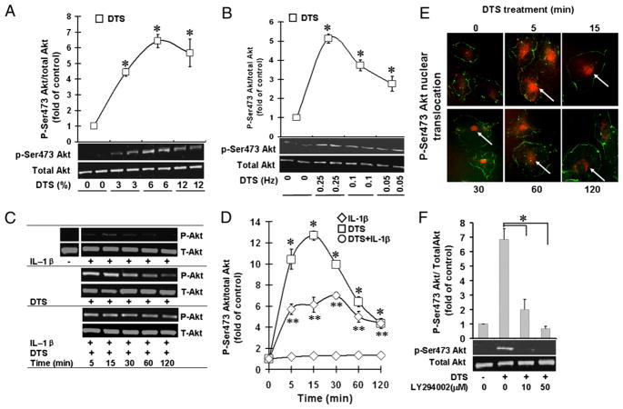
FIGURE 2.
Mechanicalstrain upregulatesAkt activation. HDMECs were exposed to DTS, IL-1β, or DTS and IL-1β to assess the Akt phosphorylation by Western blot analysis at various magnitudes of DTS at 0.25 Hz (A); at various frequencies of DTS at a magnitude of 6% (B); during various time intervals at a magnitude of 6% and 0.25 Hz (C) and relative phosphorylation of Ser473 by digitization of phosphorylated bands (D). E, Analysis of phospho-Ser473-Akt by immunofluorescence showing its cytoplasmic and nuclear translocation, indicated by arrows, between 5 and 120 min of DTS exposure (original magnification ×400). F, Western blot analysis showing Ser473-Akt phosphorylation in the presence of PI3K inhibitor LY294002. All experiments were performed in triplicates and repeated three times. *p <0.05, cells treated with DTS versus untreated control group or inhibitor-treated group; **p <0.05, cells treated with IL-1β and DTS compared with IL-1β alone.

